Imaging

Imaging Types

*hover over image for more info

Brain MRI

Brain MRIMagnetic resonance imaging (MRI) of the BRAIN is a painless, noninvasive test that produces detailed images of your brain and brain stem. An MRI machine creates the images using a magnetic field and radio waves. There is NO RADIATION associ…

Brain MRI

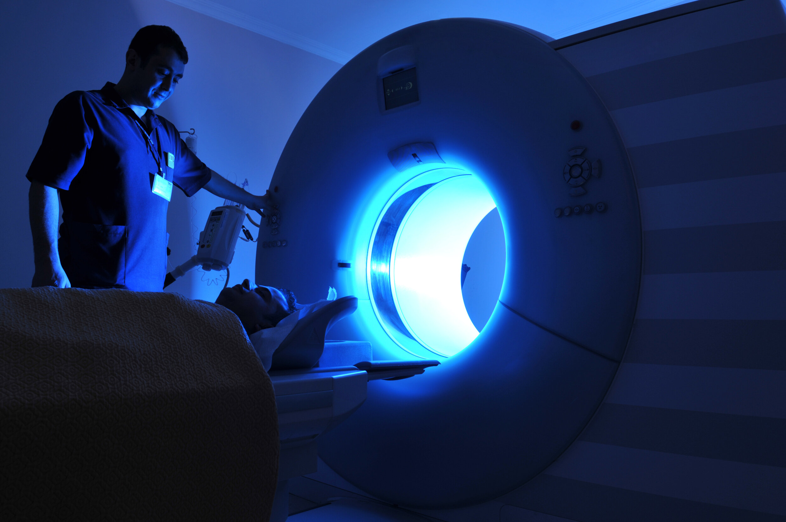
Magnetic resonance imaging (MRI) of the BRAIN is a painless, noninvasive test that produces detailed images of your brain and brain stem. An MRI machine creates the images using a magnetic field and radio waves. There is NO RADIATION associated with this detailed medical imaging modality. This test is also known as a cranial MRI.

Bone Mineral Density

Bone Mineral DensityA bone mineral density scan, also known as a DEXA scan, is a type of low-dose x-ray test that measures calcium and other minerals in your bones. The measurement helps show the strength and thickness (known as bone density or mass…

Bone Mineral Density

A bone mineral density scan, also known as a DEXA scan, is a type of low-dose x-ray test that measures calcium and other minerals in your bones. The measurement helps show the strength and thickness (known as bone density or mass) of your bones. Most people's bones become thinner as they get older. Identifying at risk individuals with low bone mineral density can allow your doctor to intervene with diet, exercise or medications that could prevent routine fractures of the spine, hip or wrist.

Brain MRA

Brain MRAAn MRA of the brain is done to look at the blood vessels leading, especially at the base of brain known as the circle of Willis, to check for an aneurysm (potentially dangerous enlargement of the blood vessel), a clot, or a narrowing (steno…

Brain MRA

An MRA of the brain is done to look at the blood vessels leading, especially at the base of brain known as the circle of Willis, to check for an aneurysm (potentially dangerous enlargement of the blood vessel), a clot, or a narrowing (stenosis) because of plaque. This imaging study does not utilize any radiation and details the vascular system of the brain and identification of a blood vessel abnormality can prevent a major vascular disorder such as a brain bleed or stroke.

MRI of the Body

MRI of the Body (Abdomen and Pelvis)MRI of the body provides clear and detailed pictures of internal organs and tissues. It has proven to be extremely valuable for the diagnosis of a broad range of pathologic conditions in all parts of the body. It …

MRI of the Body (Abdomen and Pelvis)

MRI of the body provides clear and detailed pictures of internal organs and tissues. It has proven to be extremely valuable for the diagnosis of a broad range of pathologic conditions in all parts of the body. It allows physicians to evaluate certain body structures that may not be as visible with other imaging methods.

Low Dose Chest CT

Low Dose Chest CTA CT scan of the chest is often used to further evaluate abnormalities found on routine chest x-rays. Because CT allows much greater clarity of internal organs, bone, soft tissue, and blood vessels than can be seen with routine x-ra…

Low Dose Chest CT

A CT scan of the chest is often used to further evaluate abnormalities found on routine chest x-rays. Because CT allows much greater clarity of internal organs, bone, soft tissue, and blood vessels than can be seen with routine x-rays, physicians are able to perform a more in-depth study. Preventative screening of the chest and lungs is important since lung cancers are the 3rd most common cancer in United States. Benefits: Further evaluates abnormalities found on routine chest x-rays.Help diagnose the cause of symptoms such as a cough, shortness of breath, chest pain, or fever.Evaluate an injury to the chest.Detect infections such as pneumonia, tuberculosis and COVID virus.Detect lung cancer.Demonstrate chronic conditions such as emphysema, bronchiectasis or interstitial lung disease.Diagnose vascular abnormalities such as pulmonary emboli (blood clots in the lung vessels) or an aortic aneurysm (dilated aorta).

Carotid Ultrasound

Carotid UltrasoundCarotid ultrasound is a safe, painless procedure that uses sound waves (no radiation) to examine the blood flow through the carotid arteries. Your two carotid arteries are located on each side of your neck. They deliver blood from …

Carotid Ultrasound

Carotid ultrasound is a safe, painless procedure that uses sound waves (no radiation) to examine the blood flow through the carotid arteries. Your two carotid arteries are located on each side of your neck. They deliver blood from your heart to your brain. Identifying significant narrowing of these blood vessels ahead of time may prevent a serious cerebral vascular accident (stroke).

Coronary Calcium Scan

Coronary Calcium ScanA coronary calcium scan can calculate your risk for cardiac disease and heart attack. You and your doctor can intervene before major cardiac damage or fatal heart attack occurs. Simple interventiaon such as adjusting your diet, …

Coronary Calcium Scan

A coronary calcium scan can calculate your risk for cardiac disease and heart attack. You and your doctor can intervene before major cardiac damage or fatal heart attack occurs. Simple interventiaon such as adjusting your diet, exercise and possibly medications can dramatically reduce your cardiac risks. This test might be most helpful for people who do not have heart disease but who are at medium risk for heart disease.

Prostate Ultrasound

Prostate UltrasoundUltrasound of the prostate in men uses sound waves (no radiation) to produce pictures of your prostate gland.  These images can diagnose abnormalities such as enlargement as occurs in prostate infection and frequenc…

Prostate Ultrasound

Ultrasound of the prostate in men uses sound waves (no radiation) to produce pictures of your prostate gland.  These images can diagnose abnormalities such as enlargement as occurs in prostate infection and frequency urinating or irregularity of the prostate that occurs in prostate cancer.  This study is often to investigate a nodule found during routine rectal exam,


Imaging Add-Ons


PACKAGE 1 — $2,500

MRI (abdomen), MRI (pelvis)

Coronary Calcium Scan, Chest CT (low dose)

PACKAGE 2 — $3,000

Everything in Package 1

+ Brain MRI

PACKAGE 3 — $3,500

Everything in Package 2

+ Prostrate & Carotid Ultrasound

About Imaging Types

Brain MRI

Magnetic resonance imaging (MRI) of the BRAIN is a painless, noninvasive test that produces detailed images of your brain and brain stem. An MRI machine creates the images using a magnetic field and radio waves. There is NO RADIATION associated with this detailed medical imaging modality. This test is also known as a cranial MRI.

Brain MRA

An MRA of the brain is done to look at the blood vessels leading, especially at the base of brain known as the circle of Willis, to check for an aneurysm (potentially dangerous enlargement of the blood vessel), a clot, or a narrowing (stenosis) because of plaque. This imaging study does not utilize any radiation and details the vascular system of the brain and identification of a blood vessel abnormality can prevent a major vascular disorder such as a brain bleed or stroke.

Low Dose Chest CT

A CT scan of the chest is often used to further evaluate abnormalities found on routine chest x-rays. Because CT allows much greater clarity of internal organs, bone, soft tissue, and blood vessels than can be seen with routine x-rays, physicians are able to perform a more in-depth study. Preventative screening of the chest and lungs is important since lung cancers are the 3rd most common cancer in United States. Benefits: Further evaluates abnormalities found on routine chest x-rays.Help diagnose the cause of symptoms such as a cough, shortness of breath, chest pain, or fever.Evaluate an injury to the chest.Detect infections such as pneumonia, tuberculosis and COVID virus.Detect lung cancer.Demonstrate chronic conditions such as emphysema, bronchiectasis or interstitial lung disease.Diagnose vascular abnormalities such as pulmonary emboli (blood clots in the lung vessels) or an aortic aneurysm (dilated aorta).

Coronary Calcium Scan

A coronary calcium scan can calculate your risk for cardiac disease and heart attack. You and your doctor can intervene before major cardiac damage or fatal heart attack occurs. Simple interventiaon such as adjusting your diet, exercise and possibly medications can dramatically reduce your cardiac risks. This test might be most helpful for people who do not have heart disease but who are at medium risk for heart disease.

MRI of the Body (Abdomen and Pelvis)

MRI of the body provides clear and detailed pictures of internal organs and tissues. It has proven to be extremely valuable for the diagnosis of a broad range of pathologic conditions in all parts of the body. It allows physicians to evaluate certain body structures that may not be as visible with other imaging methods.

Carotid Ultrasound

Carotid ultrasound is a safe, painless procedure that uses sound waves (no radiation) to examine the blood flow through the carotid arteries. Your two carotid arteries are located on each side of your neck. They deliver blood from your heart to your brain. Identifying significant narrowing of these blood vessels ahead of time may prevent a serious cerebral vascular accident (stroke).

Prostate Ultrasound

Ultrasound of the prostate in men uses sound waves (no radiation) to produce pictures of your prostate gland.  These images can diagnose abnormalities such as enlargement as occurs in prostate infection and frequency urinating or irregularity of the prostate that occurs in prostate cancer.  This study is often to investigate a nodule found during routine rectal exam,

Bone Mineral Density

A bone mineral density scan, also known as a DEXA scan, is a type of low-dose x-ray test that measures calcium and other minerals in your bones. The measurement helps show the strength and thickness (known as bone density or mass) of your bones. Most people's bones become thinner as they get older. Identifying at risk individuals with low bone mineral density can allow your doctor to intervene with diet, exercise or medications that could prevent routine fractures of the spine, hip or wrist.